Optical Coherence Tomography1
(OCT)
is a newly established noninvasive imaging technique which is used here for in vivo biocompatibility studies of percutaneous2 implants.
A drawback of this technique is that the OCT scans are optical distorted. The distortion mostly depends on the optical refraction within the x-rayed material.
Refraction correction of the OCT scans is an essential step towards further tasks like morphometric analysis of the implant--tissue interface.
As modern OCT scanners are capable for near realtime image capturing with high image resolutions and therefore generate huge amounts of data, methods for automatic image processing are required.

|

|
|---|---|
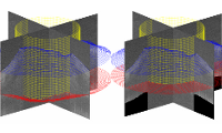
| Figure 1: Model of a percutaneous implant and volume rendering of a skin tissue (the hole indicates the position of the implant pin) | Figure 2: 3D segmentation (blue: skin surface, red: base contour, yellow: pin) and refractive undistortion |
The goal of this project is to provide a fully automatic refractive undistortion framework of the OCT scans using model based 3D segmentation.
The first step involves the segmentation of the skin and percutaneous implant base using Markov random fields and a generalized Hough transform approach.
In the second step, the amount of image distortion is estimated using a refractive distortion model.
Model parameters are estimated by incorporating a-priori knowledge of the planar shape of the percutaneous implant base.
Finally, the images are undistorted according to the refractive distortion model and the previously estimated model parameters.
