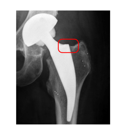
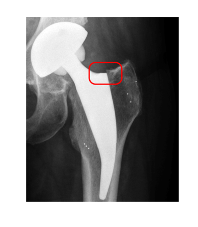
Each year more people have to live with an artificial joint. Especially the numbers of hip and knee replacements are growing recently. Additionally, the average age for the initial implantation is decreasing and patients have to live longer with their replacement. Often at least one revision is needed in a patient's life time, with each operation destroying more healthy bone tissue.
Therefore, orthopedists and implant manufacturers aim for prostheses with a long service life of more than 15 years. Objective measurements are essential to evaluate newest developments in implant design and fixation techniques. Highly accurate methods that measure the amount of implant migration, i.e. the movement of the implant relative to the surrounding bone, significantly reduce sample size as well as study length. Detected micromotions in the first two years correlate with early loosing of the implant and are thus an established measurement for quality.


Roentgen Stereophotogrammetric Analysis (RSA) is the current method of choice for evaluating implant migration. The use of two X-ray tubes at the same time allows for the reconstruction of the three-dimensional scene pictured in the two-dimensional images. Tantalum markers attached to the implant and inserted into the bone can be detected easily and accurately in the images. Since the images are calibrated the three-dimensional positions of the markers can be triangulated and used to compute the rigid motion between each follow-up examination. Thus, the migration over time can be determined.
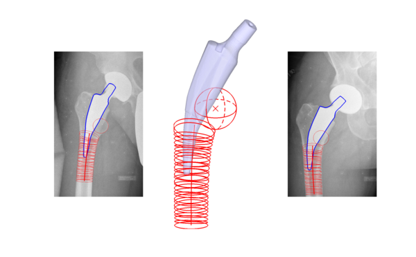
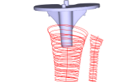
Model-based approaches use well-known three-dimensional models of the implant and the bone instead of markers to estimate the pose, i.e. location and orientation, of the models given the two-dimensional images.
The marker-based RSA has several drawbacks related to the use of the metal markers and the stereo setup. A marker-less method working in a monocular imaging setup with comparable accuracy is the final goal of the research at the TNT. The focus lays on automation of existing methods and the development of algorithms that work solely on the two-dimensional images without the need of three-dimensional patient data like CT- or MRI-images.

Research is carried out in cooperation with the Laboratory for Biomechanics and Biomaterials of the Hannover Medical School.
